Roche IRISe Explorer Diagnostics R&D Platform
Accelerating medical decisions with AI
IRISe Explorer is a digital healthcare imaging platform that uses AI to accelerate medical image review for patient diagnosis. I enhanced the image viewer to streamline experts’ review across multiple image types, helping expand the platform’s adoption within the radiology community.
Role
UX Designer
Team
Product managers (Technical, Business), Engineering teams (Frontend, Backend, Quality assurance)
Duration
Oct ~ December 2023 (2 months)
Impacts
User acquisition (Radiology institution),
Design pattern creation for company-wide design system
-
Transformative list view for focused validation
Analyzing medical images involves taking a close look at organs or cells and comparing them with one another. This critical task requires deliberate human validation. To assist physicians in validation, I enhanced the list design that contains all cell and organ annotations. At the highest level, it displays items vertically for easy skimming and converts to a horizontal layout for more detailed inspection.
-
Optimized navigation for sequential image review
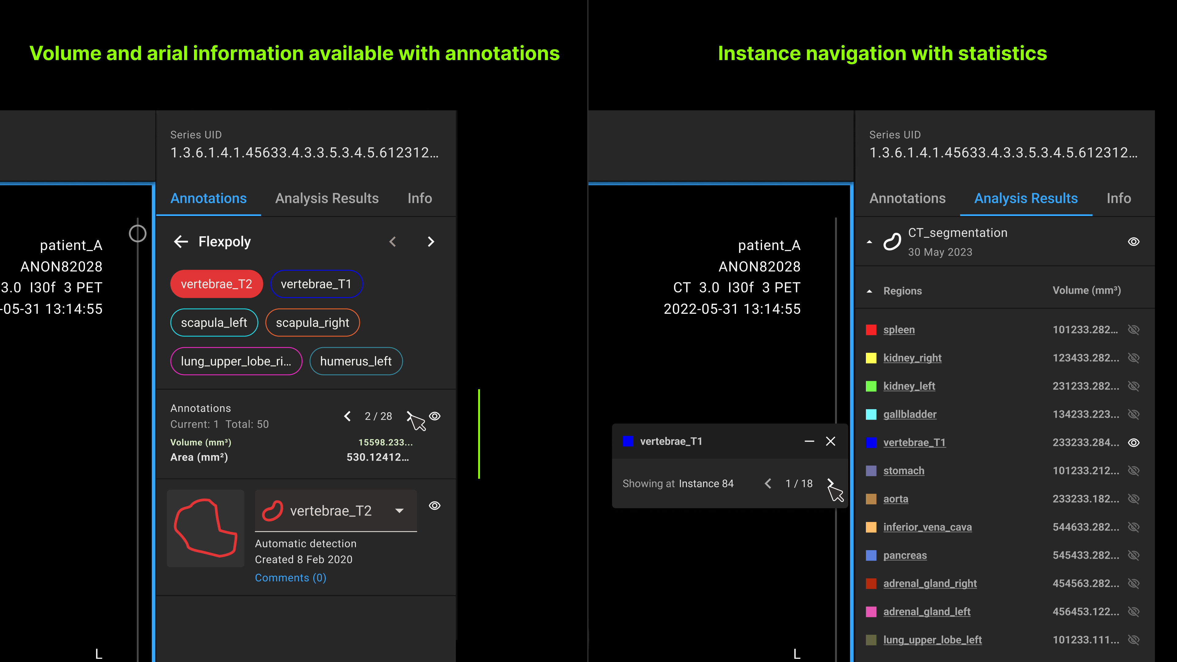
Finding insights in a patient’s image can be daunting, Navigating through hundreds of image slices, such as in a CT scan, increases the risk of missing small annotations that could signal a health issue. To mitigate this risk, I designed a new navigation system that links to all annotations of interest across multiple images. This allows radiologists to more efficiently track the trend of annotations, improving their ability to identify potential issues.
-
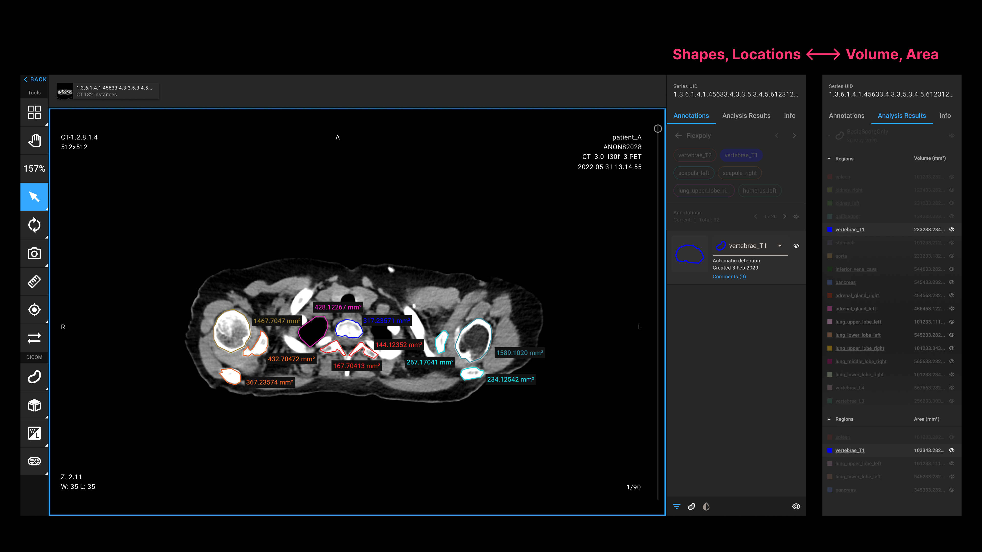
Easy referencing for insight discovery
During the image review, radiologists often refer to various information to make the most accurate diagnosis. However, this information is scattered, which can be distracting. To streamline the process, I improved the accessibility of the necessary information. No matter which menu radiologists are at, they can confirm the statistical information of all annotation to identify severe symptoms.
How does AI accelerate medical decisions?
Have you ever wondered why it takes so long to receive your pathology test results? It’s because many of the processes are done manually, and pathologists have to make annotations all by themselves while reviewing tissue samples.
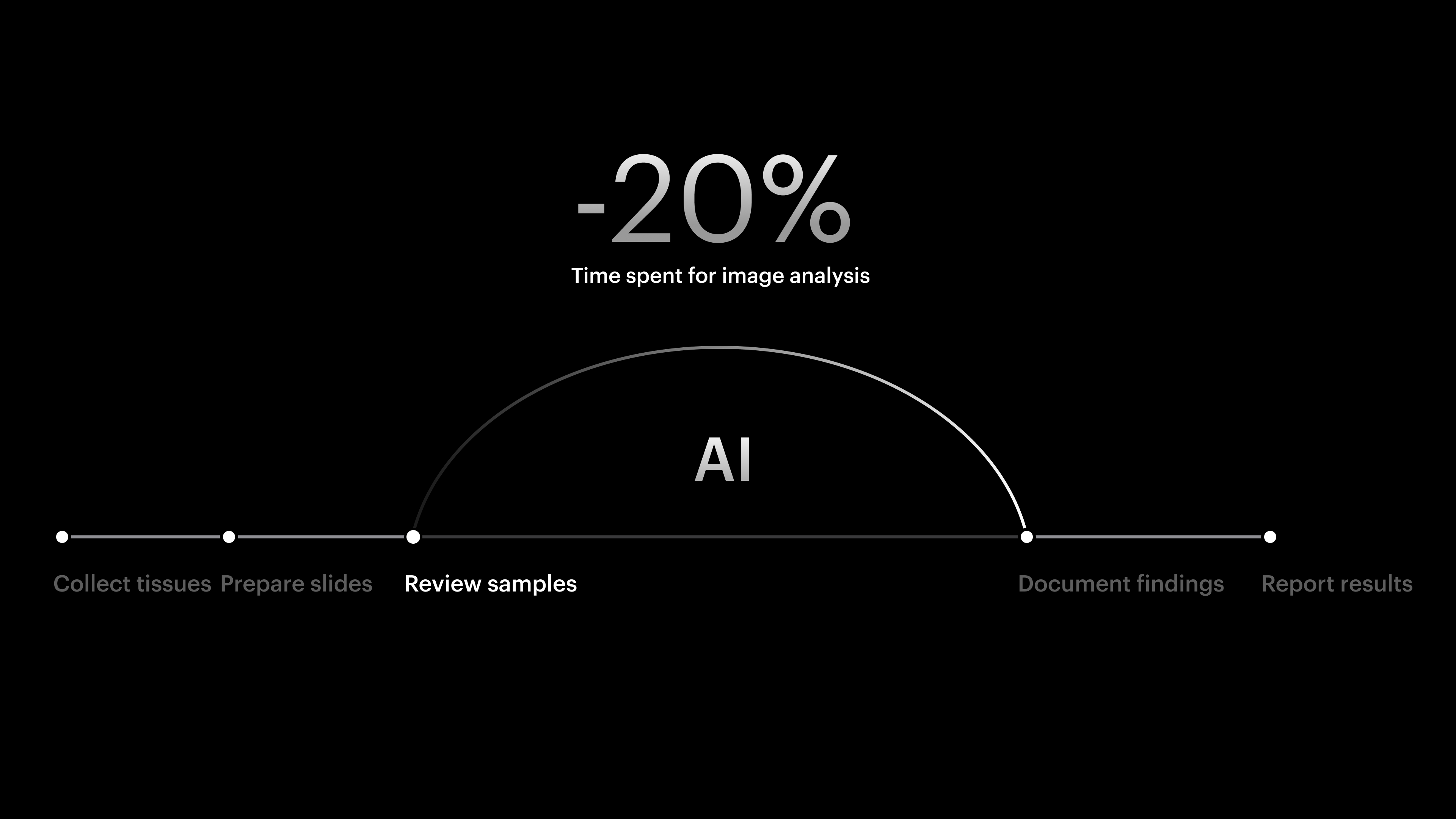
All of this can be sped up by using AI for the review process. With AI, pathologists can generate annotations even before they start reviewing, saving up to 20% of the time spent on image analysis. This means clinicians can provide treatment to patients 20% faster.
How might we scale up the product while satisfying our existing advanced users?
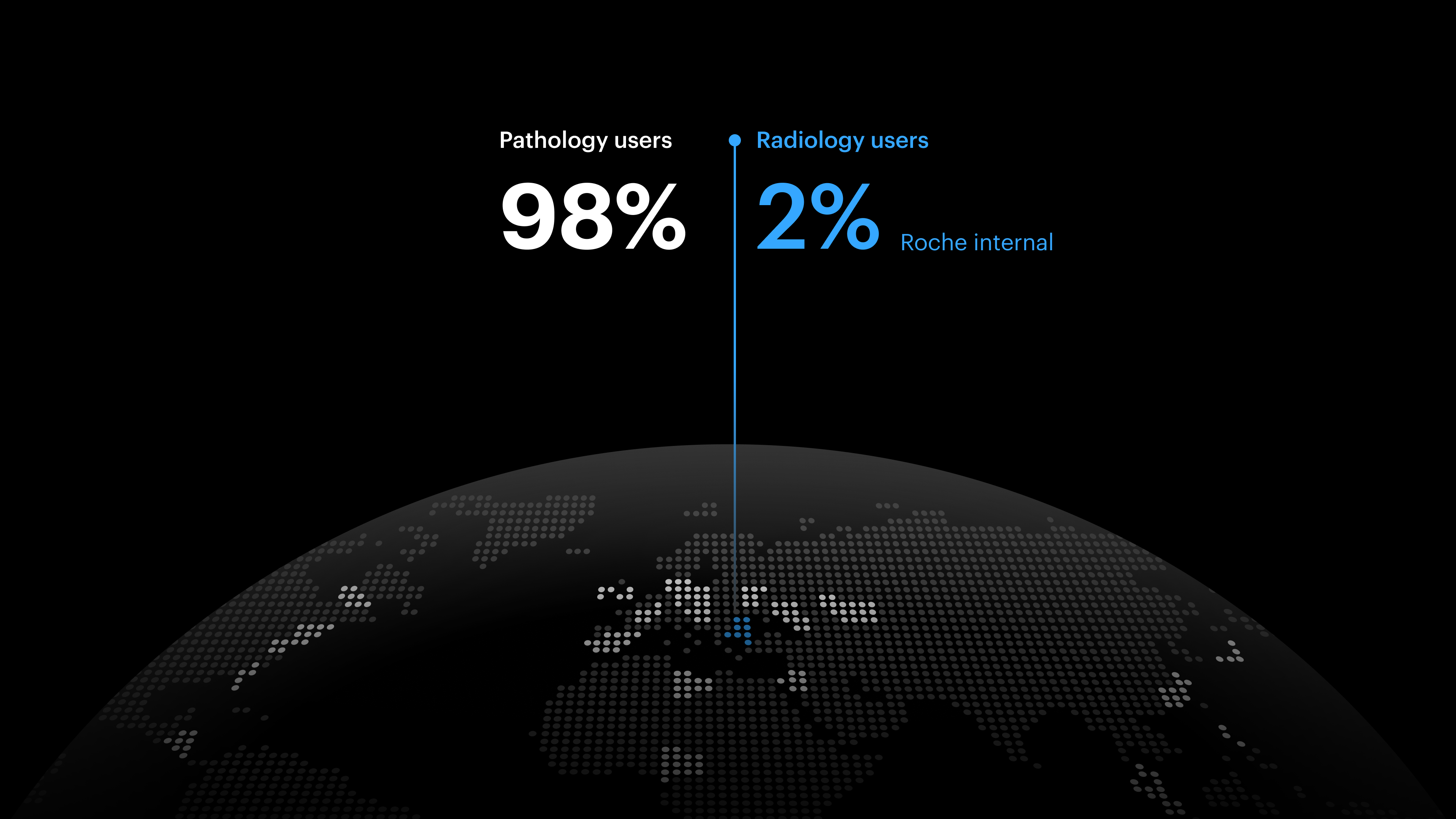
The biggest challenge in IRISe’s product growth was that it had dedicated user base. As IRISe has primarily served pathology users, all of the experience was tailored to pathology use cases. Since most of them are heavy users actively using IRISe, even subtle change could affect their current experience and cause confusion. This was a serious blocker to the product expansion into radiology.
Identifying the growth opportunity that mutually benefits advanced and potential users
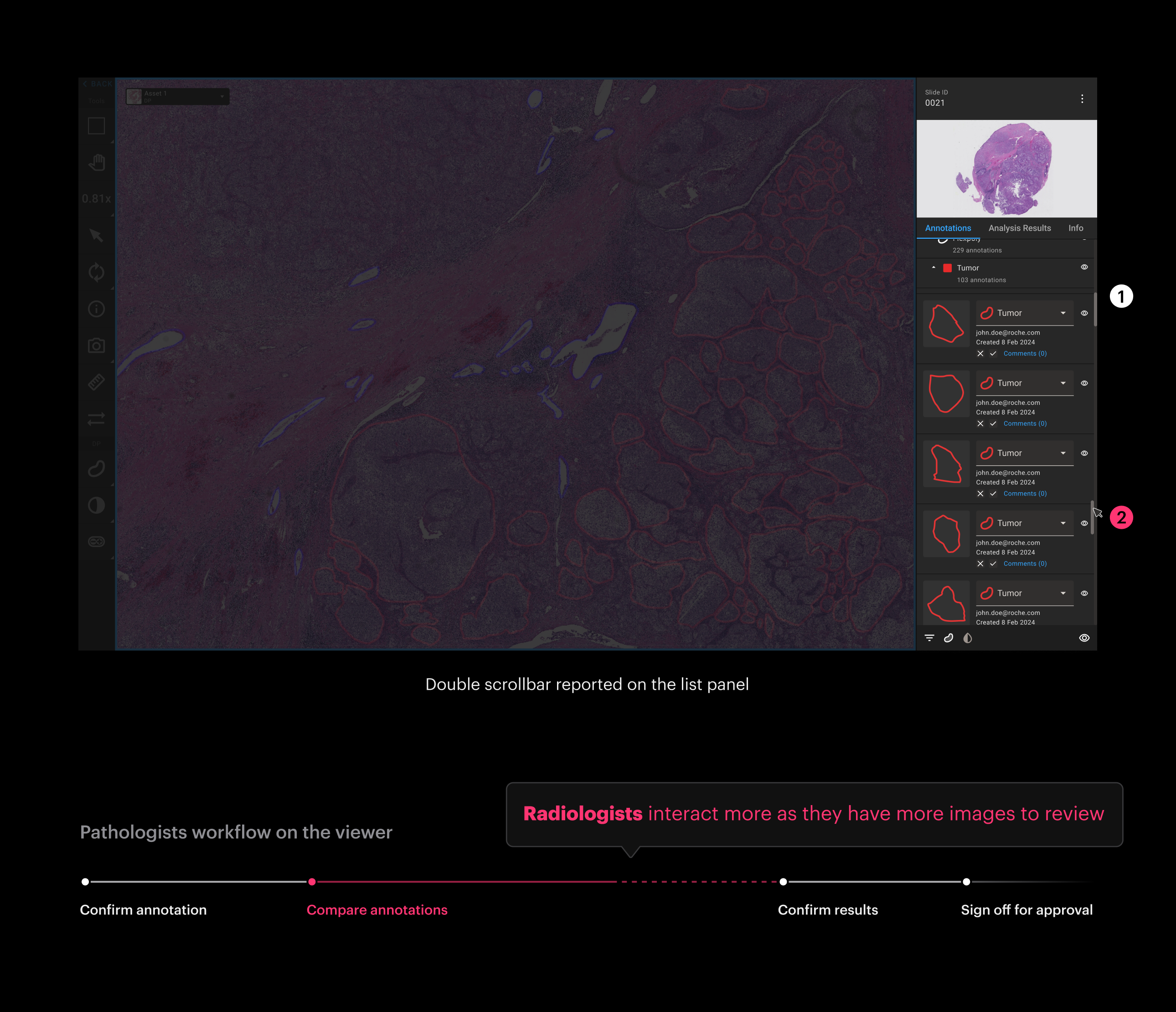
This all started with a bug report. There was a bug fix reported for the image viewer, where our users spend most of their time. As it being the most interacted area, I saw it as an opportunity to initiate the design for our product’s expansion into radiology.
Analyzing the workflows of both pathologists and radiologists, I identified design opportunities that support the primary tasks of both groups and convinced the team to work on this enhancement.
Comparable list for multimodal patient image analysis
By improving the annotation review process, I successfully introduced the new experience to our advanced and potential users. While going through multiple rounds of reviews and iterations with our users and teams, the design principles I had in mind were:
- Conciseness Prioritize streamlining the review process with intuitive navigation for complex annotation sets, reducing the learning curve for existing users
- Scalability Build a flexible design foundation to accommodate additional image formats beyond pathology and radiology, enabling advanced analysis
- Adaptability Elevate the design pattern across Roche’s research decision support portfolio to enhance UX consistency and improve implementation efficiency
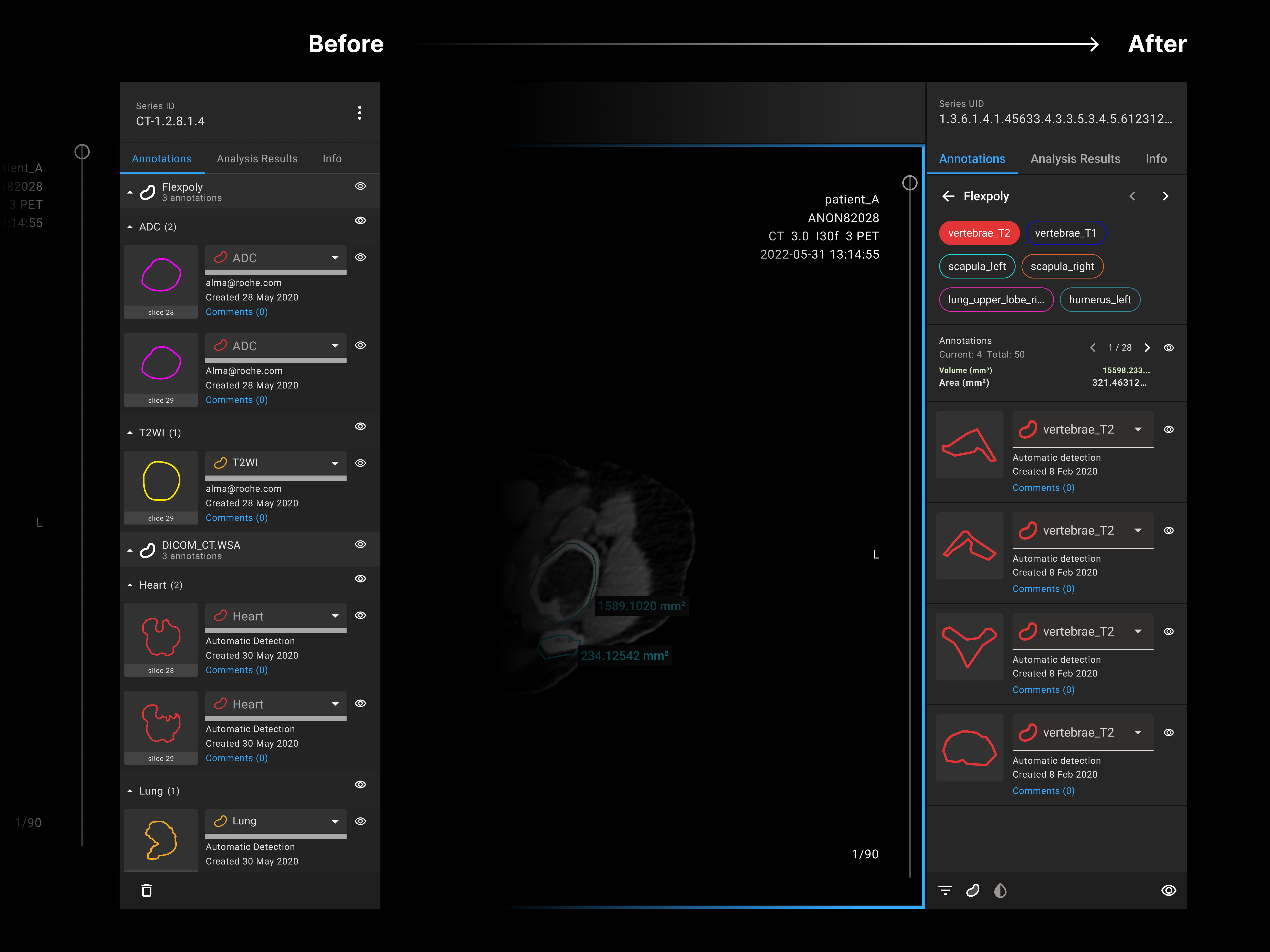
Designing for new users
Scaling the product to a new domain entailed viewing the product from different angles and addressing edge cases. By inviting targeted users from radiology, I uncovered their unique needs, which guided my design iterations.
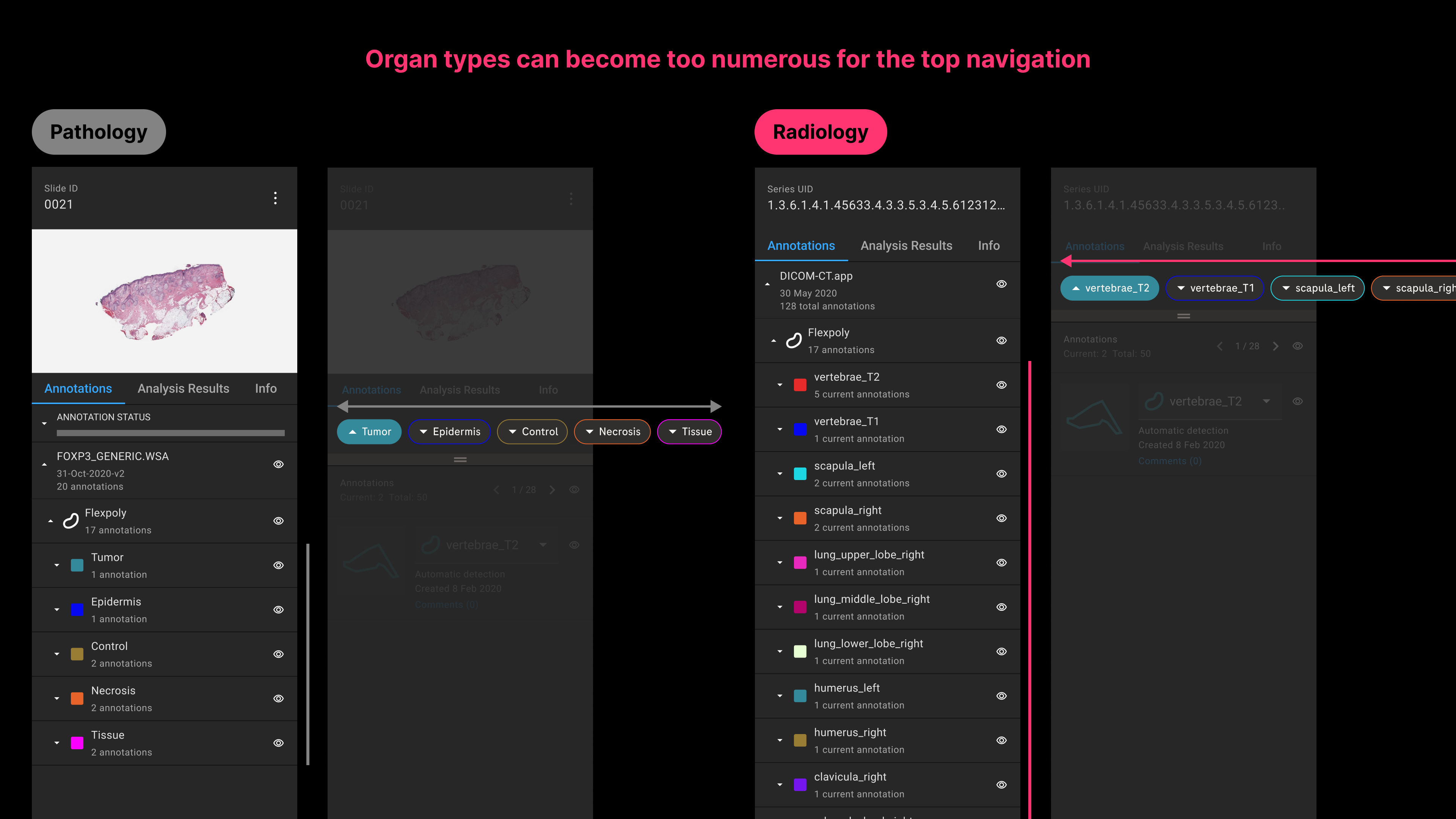
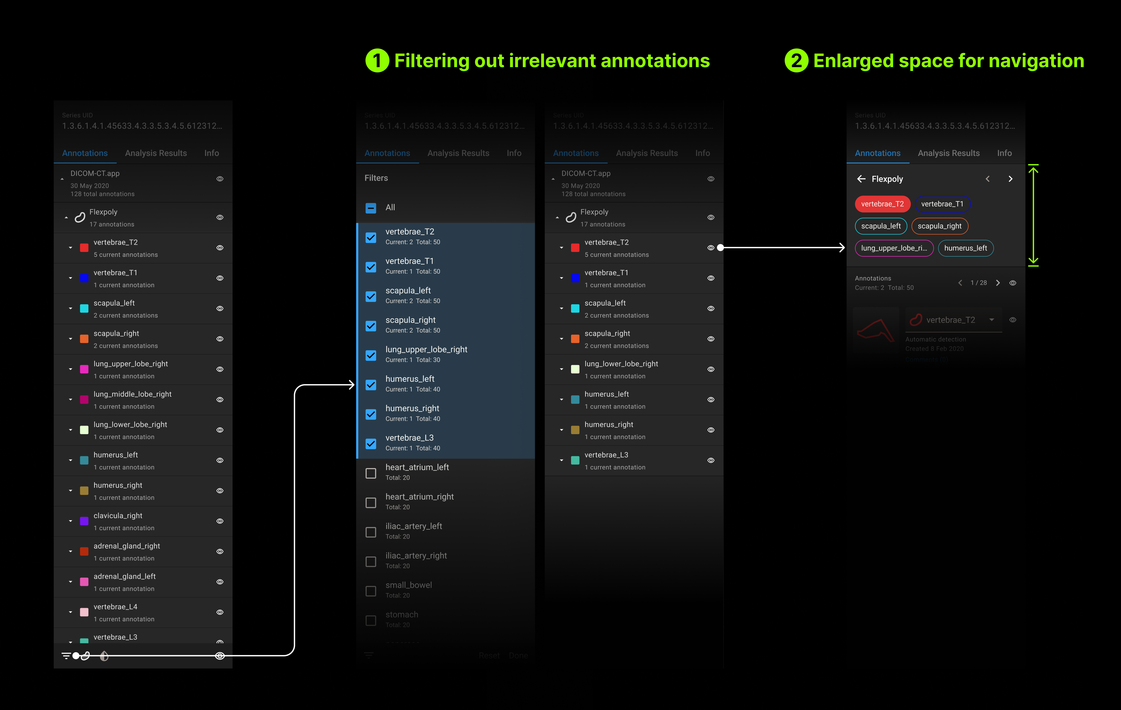
Unlimited AI capacity affecting user actions
Generating annotations with AI reduces significant amount of work for radiologists, but it can become unmanageable if too many are generated. From my conversations with Radiologists, I learned that they have a AI solution that generates annotations for all human organs. Knowing this, I iterated on my initial navigation design to make it more navigable.
More data for insights
Radiologists have more images to review than others as the image is comprised of a series of image slices. To find insights from these images, they wanted to confirm statistical data and identify the largest annotation of interest. This caused them to navigate back and forth between different menus to confirm the information, which distracted them from the review. To address this, I made those information more accessible in both contexts.
Multi-image viewer utilizing multiple AI algorithms
The ultimate goal of the enhancement was to enable clinical experts to leverage both pathology and radiology data to obtain more precise insights. Now, the viewer supports viewing multiple images on a panel, where users can compare outcomes and write reports. The seamless transition between different image analyses ensures that users can focus on insights with no distractions.
After the new viewer release, my team and I were able to observe the positive impacts such as:
Onboarding the new radiology users. This was a substantial step forward for IRISe as they committed to developing algorithms for radiology during their research, which could also be applicable for other radiology users. The power of technology!
Opening up the possibilities for related functionalities on IRISe. Previously, due to the less flexible viewer design, some advanced functionalities were disregarded by our users. With the enhanced experience, we received positive feedback from users, shedding light on how we should move forward.
Publishing the new design pattern for other products. The new list view has been adopted by other products, greatly increasing our UX consistency and reducing technical costs.